• Products
  • Solutions
  • Support
  • Contact
  • Order OnlineOrder Online

Top 10 Bone Biopsy Procedure Questions Answered?

A Bone Biopsy Procedure can turn an uncertain scan into a clearer clinical decision. It may identify infection, benign growth, metastatic disease, or a primary bone tumor. The process usually involves imaging review, needle placement, tissue collection, and laboratory analysis. Small details matter, including the entry point, specimen depth, and communication with the pathologist.

The International Agency for Research on Cancer estimated 20 million new cancer cases worldwide in 2022. However, primary malignant bone tumors remain uncommon, representing less than 1% of all cancers. The National Cancer Institute also emphasizes that imaging alone cannot reliably distinguish every bone lesion. Tissue confirmation is often necessary. Dr. David M. Panicek, a leading musculoskeletal radiologist at Memorial Sloan Kettering Cancer Center, has emphasized, “The biopsy should be planned as carefully as the definitive surgery.” That principle deserves attention.

Planning prevents avoidable problems.

This guide answers ten practical Bone Biopsy Procedure questions, including preparation, imaging guidance, pain control, sample quality, complications, recovery, and pathology timing. It also explains why the safest biopsy route should be coordinated with the orthopedic oncology team. A poorly positioned tract can complicate later surgery, although modern image-guided techniques have improved precision. No pathway is flawless. Patient anatomy, lesion location, infection risk, and operator experience can change the plan. Readers should also remember that published statistics describe groups, not individual outcomes. Clear questions remain valuable, especially when a scan looks alarming but the diagnosis is still uncertain.

Top 10 Bone Biopsy Procedure Questions Answered?

What Is a Bone Biopsy and When Is It Recommended?

A bone biopsy removes a small sample of bone or nearby tissue for laboratory examination. Doctors use imaging to guide a needle through the skin in many cases. This is called a needle biopsy. An open biopsy requires a small surgical incision. The tissue is examined by a pathologist under a microscope. It may reveal infection, an unusual bone lesion, or cancer cells.

A biopsy is recommended when scans show an area that remains unclear or suspicious. It can also confirm whether a known disease has reached the bone. A musculoskeletal radiologist or orthopedic surgeon considers the lesion’s location, size, appearance, and the patient’s symptoms. The choice is not always obvious. Sometimes, repeat imaging is safer than immediate sampling. Other times, delaying the biopsy could postpone essential treatment.

Before the procedure, patients should discuss blood-thinning medicines, allergies, and previous operations. Local anesthesia is common, while deeper lesions may require sedation. Temporary soreness or bruising can occur afterward. Pain should be reported if it becomes severe, persistent, or comes with fever. Results may take several days because special laboratory tests are sometimes needed. Occasionally, the sample is too small or does not explain the scan. A second biopsy may then be considered, which can feel frustrating but may improve diagnostic accuracy.

How Should Patients Prepare for a Bone Biopsy?

How Should Patients Prepare for a Bone Biopsy?

Preparation starts with an honest medication review. Tell the care team about blood thinners, diabetes medicines, allergies, supplements, and pregnancy. Do not stop prescribed medicine alone. The American College of Radiology and Society of Interventional Radiology recommend checking bleeding risk before image-guided biopsy. Your clinician may request blood tests, including platelet count and clotting studies. Results can change the procedure plan.

Fasting depends on sedation. The American Society of Anesthesiologists advises avoiding solid food for about six hours before elective sedation, while clear liquids may be allowed until two hours beforehand. Follow your hospital’s instructions instead. Wear loose clothing and arrange transport if sedation is planned. Bring scans, medication lists, and a responsible adult. Pain is usually controlled, but experiences differ. I would not assume a “small” biopsy feels simple for every patient.

Tips: Ask whether local anesthesia, sedation, or both will be used. Confirm when you should stop eating and drinking. Leave jewelry at home. Tell staff about fever, skin infection, or new illness. Afterward, keep the dressing clean and watch for increasing redness, drainage, fever, or uncontrolled pain. The National Cancer Institute notes that biopsy results can require additional testing, so ask when and how results will arrive. That waiting period is often harder than expected.

What Happens During the Bone Biopsy Procedure?

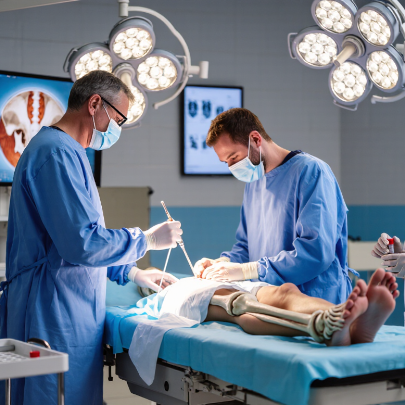
A bone biopsy usually begins with a review of scans, medical history, medicines, and allergies. The clinician explains the approach and obtains informed consent. Most percutaneous biopsies use local anesthetic, while some patients receive light sedation. You may feel pressure, but sharp pain should be reported immediately. The area is cleaned carefully, then a biopsy needle passes through the skin. CT, X-ray, or ultrasound guidance helps target the abnormal bone accurately. Several small tissue samples may be collected. A dressing covers the entry point afterward.

A systematic review in European Radiology reported diagnostic yields above 90% for image-guided bone biopsy, although results vary by lesion, location, and technique. Reported complication rates were low, but bleeding, infection, nerve irritation, or an inadequate sample can still occur. “Minimally invasive” does not mean completely painless. That wording sometimes creates unrealistic expectations. The specimen then travels to pathology, where specialists examine cells and may order additional tests. Results can take several days or longer.

Tips: Wear loose clothing and follow fasting instructions if sedation is planned. Tell the team about blood thinners, diabetes, and previous anesthesia problems. Arrange transportation if sedatives are used. Afterward, keep the dressing clean and watch for increasing pain, fever, swelling, or bleeding. Contact the care team promptly if these appear. A missed detail in your medical history can change preparation, so honest answers matter.

Top 10 Bone Biopsy Procedure Questions Answered? - What Happens During the Bone Biopsy Procedure?

No. Common Question What Usually Happens Key Considerations
1 Why is a bone biopsy performed? A bone biopsy removes a small sample of bone or tissue for laboratory examination. It may help determine whether an abnormal area is caused by infection, a benign condition, or cancer, and can help guide treatment. Imaging tests can show where an abnormality is located, but a biopsy may be needed to identify its exact nature.
2 What are the main types of bone biopsy? A needle biopsy uses a hollow needle to collect one or more samples through a small skin opening. An open biopsy uses a surgical incision to expose and remove a larger sample of bone. The method depends on the location, size, and appearance of the lesion, the suspected diagnosis, and the amount of tissue required.
3 How should a patient prepare? The medical team reviews medical history, allergies, medicines, and previous imaging. Blood-thinning medicines may need special instructions. Fasting may be required if sedation or general anesthesia is planned. Patients should not stop prescribed medicines or eat, drink, or drive restrictions without specific instructions from the care team.
4 What happens before the biopsy begins? The patient is positioned so the target bone is accessible. The skin is cleaned with an antiseptic solution, and imaging such as CT, ultrasound, or X-ray may be used to plan and guide the biopsy. The planned approach is selected to reduce injury to nearby nerves, blood vessels, and other tissues.
5 Will the procedure be painful? Local anesthetic is commonly injected to numb the area. Depending on the biopsy site and patient needs, sedation or general anesthesia may also be used. Pressure or pushing may be felt even when sharp pain is controlled. Temporary soreness, bruising, or tenderness around the biopsy site is common after the procedure.
6 How is a needle biopsy performed? After a small skin incision, the clinician advances a biopsy needle through the soft tissue and into the abnormal bone, often using imaging guidance. One or more tissue or fluid samples are collected and sent for analysis. A dressing or small bandage is usually placed over the entry site after the needle is removed.
7 How is an open bone biopsy different? During an open biopsy, a surgeon makes an incision, exposes the bone, removes a tissue sample, and closes the incision with sutures or other wound-closure materials. It generally involves more wound care and recovery than a needle biopsy and may be performed in an operating room.
8 How long does a bone biopsy take? The biopsy itself may take approximately 30 minutes to 1 hour for many image-guided procedures, but preparation, anesthesia, observation, and recovery can make the overall visit several hours. The exact time varies according to the biopsy type, body location, imaging method, and anesthesia plan.
9 What are the possible risks? Potential risks include bleeding, infection, pain, bruising, injury to nearby structures, an inadequate sample, and—depending on the bone and procedure—weakening or fracture of the bone. Serious complications are uncommon, but individual risk depends on health conditions, medicines, lesion location, and biopsy technique.
10 What happens after the biopsy? The patient is monitored until vital signs are stable. Instructions may include keeping the dressing clean and dry, limiting strenuous activity, and using recommended pain relief. A pathologist examines the sample, and results may take several days or longer. Contact the healthcare team for worsening pain, persistent bleeding, fever, increasing redness or swelling, drainage, numbness, or difficulty moving the affected area.

Information is general and does not replace individualized advice from a qualified healthcare professional.

What Risks and Recovery Issues Should Patients Expect?

What Risks and Recovery Issues Should Patients Expect?

Bone biopsy usually involves removing a small tissue sample with a needle or surgical incision. The approach depends on the bone’s location, imaging findings, and suspected condition. In a large review published in the Journal of Vascular and Interventional Radiology, image-guided bone biopsy achieved diagnostic yields of roughly 90–95%. Serious complications were uncommon, but they were not impossible.

Pain, bruising, minor bleeding, and temporary swelling are the usual recovery concerns. Some patients return home the same day, especially after a needle biopsy. Local soreness may last several days. Surgical biopsies can require longer restrictions, wound care, and help with movement. The American College of Radiology and Society of Interventional Radiology emphasize individualized planning, including medication review and bleeding-risk assessment.

Watch the biopsy site carefully.

Increasing redness, warmth, drainage, fever, severe pain, numbness, or uncontrolled bleeding requires prompt medical advice. Rarely, infection, nerve irritation, fracture, or an inadequate sample can occur. A failed sample may mean repeating the procedure, which is frustrating and emotionally difficult. “Minimally invasive” should never mean risk-free. Recovery also varies with bone strength, diabetes, anticoagulant use, and the biopsy location. Patients should ask when to resume driving, exercise, bathing, and weight-bearing. Advice from online sources can be too general. The treating team’s instructions matter more.

Bone Biopsy: Typical Procedure and Recovery Timeline

Most image-guided bone biopsies are completed in about 30–60 minutes, although preparation and observation may take longer. Mild pain or bruising commonly improves within 1–3 days. Light activities can often resume within 1–2 days, while strenuous activity may need to be avoided for approximately 3–7 days. Pathology results commonly take 2–7 days, depending on the tests required. Actual risks and recovery times vary by biopsy site, technique, medical condition, and clinician instructions.

How Are Bone Biopsy Results Interpreted and Used?

Bone biopsy results are interpreted as part of a larger clinical picture. A pathologist examines tissue architecture, cell appearance, and the relationship between abnormal cells and bone. The report may identify a benign lesion, primary bone cancer, metastatic disease, infection, or a non-diagnostic sample. Imaging remains essential. MRI shows local structure, while CT can reveal mineral patterns and cortical damage.

The World Health Organization’s Classification of Tumours, 5th edition, recommends integrating morphology, immunohistochemistry, and molecular findings for difficult bone tumors. These tests can separate look-alike conditions. A genetic result alone does not establish a diagnosis. It must match the microscope findings and clinical history. The National Cancer Institute’s SEER Cancer Stat Facts notes that bone and joint cancers account for less than 1% of new cancers. Their rarity makes specialist review especially valuable.

Results guide surgery, drug treatment, radiation planning, or observation. They may also determine whether another biopsy is needed. Sampling error is real. A needle may collect inflammation beside a tumor, or miss a mixed lesion entirely. That part is uncomfortable.

Doctors often compare the pathology report with scan findings and laboratory results. If they conflict, a multidisciplinary review can prevent rushed decisions. Patients should ask about the exact diagnosis, test limitations, and whether expert second review is appropriate. Even a confident report deserves context. Mistakes can happen.